OCT in Ophthalmology
Optical coherence tomography (OCT) is a fundamentally new biomedical imaging technology that generates high-resolution, cross-sectional and volumetric image of subsurface tissue structure and pathology by measuring echo time delays of light. OCT was developed by David Huang when he was an MD-PhD student at Harvard-MIT [1]. OCT performs optical biopsy however images can be obtained in real time, without the need to excise specimens. The technology has become a standard of care in ophthalmology, and over 30 million imaging procedures are performed worldwide every year [2]. It is also a powerful tool for fundamental research and pharmaceutical development. A recent study estimates that OCT has saved 9 billion dollars in healthcare costs in the US for the treatment of age related macular degeneration [3].
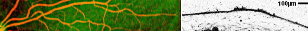
Our group’s primary focus is on developing new optical imaging technology and processing strategies to study disease pathogenesis and investigate markers of disease progression. We are interested in developing the next generation of OCT technology (Figure 1) using ultrahigh speed swept-source OCT (SS-OCT) systems for OCT angiography (OCTA) imaging of the retinal microvasculature (Figure 2) and ultrahigh resolution spectral-domain OCT (SD-OCT) systems for imaging the retina’s fine structural details (Figure 3). Changes in microvasculature or structure are early marker of disease and investigations are important for understanding pathogenesis and developing therapies.

Figure 1. Optical coherence tomography (OCT) used advanced laser sources, fiber optic interferometer design, patient interface design, high-speed detection and signal processing. Many of these techniques are similar to coherent optical communication. The schematic shows a swept source (SS-OCT) system which uses a frequency scanned, narrow band laser to perform interferometric measurements of light echoes delays. The interference signal is detected and digitized at high speed (GSPS), then processed and Fourier transformed to obtain spatial information. Spatial information is encoded in frequency, analogous to MRI. The optical beam is scanned in order to acquire cross-sectional or volumetric images.

Figure 2. Top: Optical coherence tomography angiography (OCTA) works by acquiring repeated OCT B-scans from the same retinal location in rapid succession. In the time between the repeated OCT B-scans, blood cells (e.g., erythrocytes) travel a small distance, which manifests as a change in the speckle pattern at that location on the OCT B-scan. Using signal processing techniques this changing speckle pattern can be extracted, resulting in an OCTA image of the ocular microvasculature. Leveraging our ultrahigh speed (400 kHz+ A-scan rate) swept-source OCT (SS-OCT) system, our group has developed the variable interscan time analysis (VISTA) algorithm, which allows for measurement of relative blood flow speeds, a functionality not possible with current commercially available OCTA systems. Bottom: With our clinical collaborators our group uses OCTA to study a variety of diseases, including age-related macular degeneration (red box), and diabetic retinopathy (green box).

Figure 3. Top: 12mm, high-density ultrahigh resolution spectral-domain optical coherence tomography (SD-OCT) B-scan of a healthy human retina. Ultrahigh resolution OCT allows visualization of different retinal layers (enlargements), which are not well resolved with the resolutions of current commercially available systems. EZ: ellipsoid zone; IS/OS: inner segment/outer segment junction; CIZ: cone interdigitation zone; COST: cone outer segment tip; RIZ: rod interdigitation zone; ROST: rod outer segment tip; RPE: retinal pigment epithelium; BM: Bruch’s membrane. Bottom: In collaboration with researchers at the Pattern Recognition Lab of the Friedrich-Alexander-Universität Erlangen-Nürnberg, Germany, our group has developed software methods to register and merge multiple OCT volumes into a single, motion-corrected volume with improved signal-to-noise. These methods have been successfully translated to industry, where they are currently used in several OCT instruments.
References:
[1] D. Huang, E. A. Swanson, C. P. Lin, J. S. Schuman, W. G. Stinson, W. Chang, M. R. Hee, T. Flotte, K. Gregory, C. A. Puliafito, and J. G. Fujimoto, “Optical Coherence Tomography,” Science, vol. 254, pp. 1178-1181, Nov 1991.
[2] E. A. Swanson and J. G. Fujimoto, “The ecosystem that powered the translation of OCT from fundamental research to clinical and commercial impact [Invited],” Biomed. Opt. Express 8, 1638-1664, 2017.
[3] M. A. Windsor, et al. “Estimating Public and Patient Savings From Basic Research—A Study of Optical Coherence Tomography in Managing Antiangiogenic Therapy”, American Journal of Ophthalmology, vol. 185, 115 – 122, Jan 2018.